BIOCHEMICAL AND HISTOPATHOLOGICAL EVIDENCE OF METRONIDAZOLE-INDUCED NEUROTOXICITY IN RATS
DOI:
https://doi.org/10.4314/njpr.v21i1.1Ключевые слова:
Neurotoxicity , Purkinje cells, Oxidative stress, Metronidazole, Neurotrophic factorАннотация
Background: Metronidazole-induced neurotoxicity is a recurring challenge in pharmacotherapy of susceptible infections, however pathological evidence and biomolecular involvement needs unravelling.
Objectives: this study aims to gather neuropathologic evidence and determine biomolecular involvement in metronidazole-induced neurotoxicity.
Methods: Twenty rats were assigned to 2 groups (n=10). Group 1 was given 5 mL/kg 0.5% Tween-80®, group 2 received 50 mg/kg metronidazole. The treatments were administered via oral gavage on a daily basis. On the fifteenth day, 5 rats from each group were sacrificed under halothane anesthesia, blood and the brains were excised for toxicological analysis. The remaining animals were treated for 28 days and euthanized on the 29th day and the same parameters assessed.
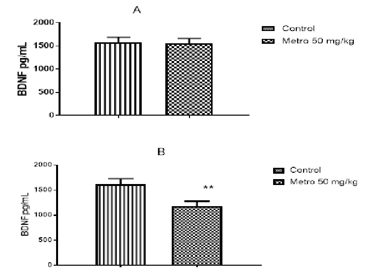
Results: Histological distortions and hemorrhage occurred in the granular layer, loss of Purkinje fibres and congestion were seen in the Purkinje layer and over expression of neuron specific enolase occurred in the cerebellum of metronidazole treated rats. Reduced superoxide dismutase activity, increased malondialdehyde (brain homogenate) and reduced serum brain derived neurotrophic factor concentrations were recorded in metronidazole treated rats.
Conclusion: Histological distortions in the granular and Purkinje layers of the cerebellar cortex, increased oxidative stress and reduced BDNF concentrations in treated rats are manifestations of metronidazole-induced neurotoxicity.
Библиографические ссылки
Загрузки
Опубликован
Выпуск
Раздел
Лицензия
Copyright (c) 2025 Nigerian Journal of Pharmaceutical Research

Это произведение доступно по лицензии Creative Commons «Attribution-NonCommercial-NoDerivatives» («Атрибуция — Некоммерческое использование — Без производных произведений») 4.0 Всемирная.
